Screening & Diagnosis

Imaging
The Imaging Unit provides diagnostic and interventional procedures to our patients. The unit is staffed with experienced radiologists, radiographers, staff nurses and other support staff who specialise in imaging services. Our top-tier facilities ensure accurate and prompt imaging services. The Diagnostic Imaging services include:
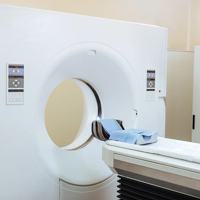
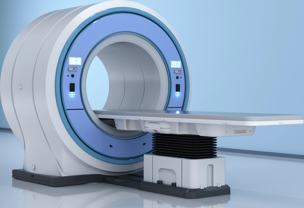
CT Scans
A CT scan, or Computed Tomography scan, is a tool used in diagnosing cancer. By providing detailed images of internal organs and structures of your body, CT scans help doctors detect abnormalities, plan treatments, and monitor the progress of therapies.
How It Works
CT scans uses X-rays and computer processing to create detailed cross-sectional images of your body. Before the scan, you will be asked to remove any metal objects or belongings. During the procedure, you will lie on a table that slides into a large, doughnut-shaped machine. As the table slides and move through the scanner, the Xrays rotate around your body taking multiple X-ray images from different angles. These images are then combined by a computer to create a comprehensive view of your internal structures. Typically, the scan lasts between 10 to 30 minutes, depending on the area being examined.
Advantages
High Accuracy and assist in monitoring disease progression
CT scans are highly effective in detecting tumours, abnormalities, and other issues within vessels, bones, organs, and tissues. A special dye known as contrast material will be given either through mouth, injection, or enema. This dye enhances the visibility of specific areas of the body being examined by appearing bright on the images.
Detailed Imaging
They provide detailed images that may guide in surgery, biopsies and the precise planning of treatments and assessment of treatment progress. CT scans can produce up to 2,000 images per scan.
Versatility
CT scans are versatile and can examine almost any part of the body, making them invaluable in cancer care.
Risks and Limitations
Radiation Exposure
CT scans involve brief exposure to radiation, and they have a higher amount of radiation compared to X-rays due to their detailed information-gathering ability. While a single CT scan has not been shown to cause long-term risks, repeated scans could pose a potential risk. However, the diagnostic benefits generally outweigh these risks.
Allergic Reactions
Although it is uncommon, some patients may have allergic reactions to the contrast materials used during the scan. Potential mild reactions include rashes or itchiness. In rare cases, the allergic reaction can be serious or even life-threatening. If you notice any signs of a reaction, please call your nearest accident and emergency, or contact the patient navigator.
Pregnancy
Inform your healthcare professional if you are pregnant
Contact WhatsApp
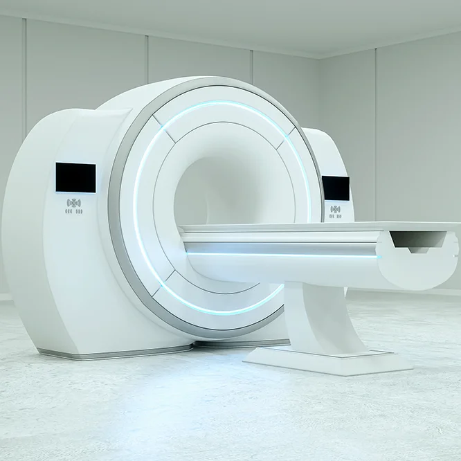
MRI Scans
Magnetic Resonance Imaging (MRI) is a non invasive imaging technique using a magnetic field and computer generated radio- waves to create detailed images of organs and tissues in the body. The detailed images produced by MRI will help and assist doctors in detection of abnormalities, plan treatments, and monitor disease progression. MRI is particularly effective in imaging soft tissues, such as the brain, spinal cord, joints, and organs. It is often used for neurological, musculoskeletal, and abdominal imaging.
How it works
Unlike CT Scans (hyperlink), MRI does not involve ionising radiation but instead uses a strong magnetic field and radio waves to generate detailed images of the body's internal structures. During the procedure, you will lie on a table that slides into a large cylindrical machine that will create a strong magnetic field around you and send pulses of waves from a scanner. The magnetic field temporarily realigns hydrogen atoms in your body, and radio waves cause these atoms to produce signals, which are used to create images. Typically, an MRI scan lasts between 30 to 60 minutes, depending on the area being examined.
Advantages
High Precision
MRIs provide highly detailed images of soft tissues, making them particularly useful for detecting tumors, brain disorders, and joint abnormalities.
No Radiation
MRIs do not use ionizing radiation, reducing potential risks associated with repeated imaging.
Contrast Material
MRI scans offers better soft tissue contrast, allowing for clearer differentiation between fat, water, muscle, and other soft tissues.
Versatility
MRI can examine almost any part of the body, including the brain, spine, joints, and organs, making it invaluable in comprehensive cancer care.
Risks and Limitations
Magnetic Field Hazards
The powerful magnetic field of MRI machines can attract metal objects and disrupt medical devices such as pacemakers. People with certain implants may not be suitable for MRI scans unless stated otherwise. It's important to inquire whether tattoos or permanent makeup might pose any issues during the procedure.
Contrast Reactions
Although it’s rare, some patients may have allergic reactions to the contrast material administered during an MRI scan.
Pregnancy and Breastfeeding
Inform your healthcare provider in advance if you are pregnant, as alternatives or postponement of the MRI may be necessary. It's also crucial to disclose if you are breastfeeding, particularly if contrast material will be used during the scan.
Contact WhatsApp
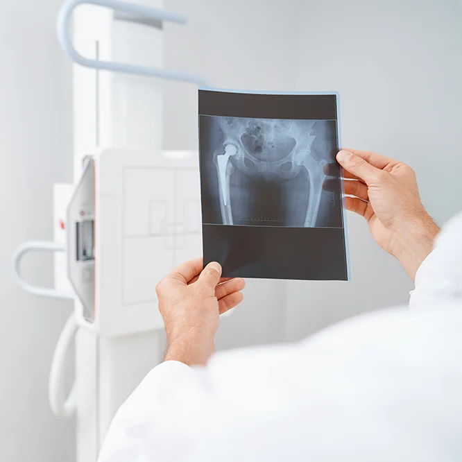
X-rays
X-rays are a form of electromagnetic radiation. In medical imaging, X-rays are directed through the body, and the resulting shadows create a two-dimensional image on a film or digital detector. X-rays are commonly used to assess bone fractures, lung conditions, and to detect abnormalities in the chest, abdomen, and other areas.
How it works
When X-rays pass through your body, different tissues absorb them at different rates. Bones, for example, absorb more X-rays and appear white on the images, while softer tissues absorb fewer X-rays and appear in shades of grey. During the procedure, you will be positioned between an X-ray machine and a special film or digital detector that captures the images. Depending on the body parts to be scanned, you will usually lie on the x-ray couch. If the area to be x-rayed is the chest, you will be told to stand or sit. The process typically takes just a few minutes.
Risks and Limitations
Radiation Exposure
X-rays involve exposure to a small amount of ionizing radiation. While the risk from a single X-ray is low, repeated exposure can increase the risk of developing cancer over time.
Image Limitations
X-rays provide less detail of soft tissues compared to other imaging techniques like MRI or CT scans.
Pregnancy Concerns
Pregnant women are advised to avoid X-rays unless absolutely necessary, as radiation can pose risks to the developing foetus.
Contact WhatsApp
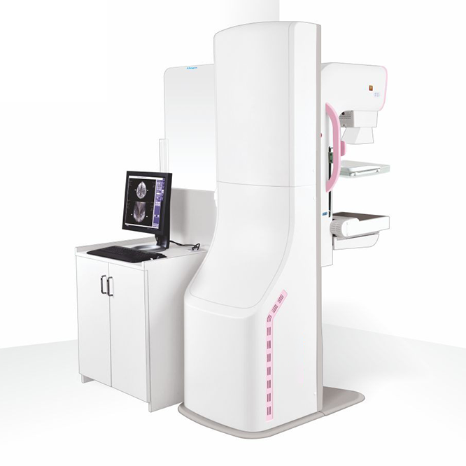
Mammography
3D Mammogram
A mammogram is an X-ray image of the breast that is often used as a tool in the early detection and prevention of breast cancer. While 2D mammograms take a single flat image, a 3D mammogram creates a three-dimensional image of the breast using X-rays by capturing multiple images of the breast from different angles. These images are then reconstructed into a 3D view, allowing radiologists to examine breast tissue layer by layer.
How It Works
During the procedure, you will stand in front of an X-ray machine, and each breast will be compressed between two firm plates to spread out the breast tissue. The machine takes multiple images from different angles, which are then compiled into a 3D image by a computer. This process usually takes about 20-30 minutes and provides a more comprehensive view compared to traditional 2D mammograms. After the exam, a radiologist will analyze the images and send a report to your healthcare provider. If any abnormalities are detected, you may be asked to return for additional imaging or a biopsy. Remember, being called back does not necessarily mean you have cancer; it often means more information is needed.
Benefits
Improved Accuracy
3D mammograms have a higher accuracy rate in detecting breast cancer compared to 2D mammograms. This is because the detailed images allow radiologists to distinguish between benign and malignant areas more effectively.
Reduced Need for Follow-Up Imaging
With clearer images, 3D mammograms reduce the likelihood of false positives, meaning fewer women need to return for additional imaging.
Better Detection in Dense Breast Tissue
Women with dense breast tissue benefit significantly from 3D mammograms, as the technology provides a clearer view of overlapping tissue structures.
Who Should Get a 3D Mammogram?
3D mammograms are recommended for all women who need routine breast cancer screening, typically starting at age 40. Women with a higher risk of breast cancer, such as those with a family history or genetic predisposition, should discuss earlier and more frequent screenings with their healthcare provider.
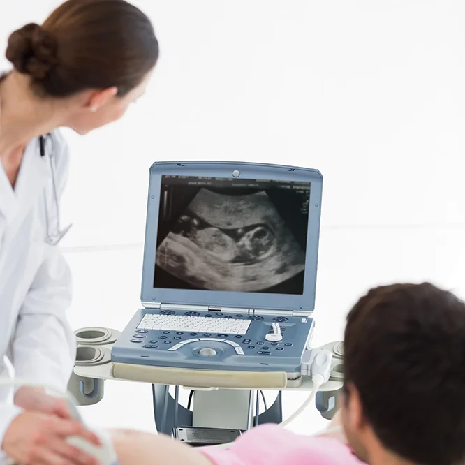
Ultrasound
Ultrasound, also known as sonography, is a common and safe imaging technique used by doctors to see inside your body. Ultrasound uses high frequency sound waves to create pictures inside your body and the structures visualised will guide to making a diagnosis and facilitate treatment . A small device called a transducer sends out sound waves and picks up the echoes as they bounce back from your internal organs.
How It Works
Depending on the area that needs to be examined, you will usually be asked to remove clothing from that part of your body and lie down on an examination table. The sonographer will then apply a thin gel layer to your skin over the area to be examined so that the ultrasound waves are transmitted from the transducer through the gel and into your body. The probe converts electrical current into high frequency sound waves and sends te waves into the body's tissues and the sound waves bounce off structures inside your body back to the probe which converts the waves into electrical signals which will be captured onto a computer screen as real-time videos or images. The procedure is painless, though you might feel slight pressure from the transducer. Once the imaging is complete, the gel will be wiped off, and you can get dressed and resume normal activities.
Risks and Limitations
Image Quality
Sometimes, images may not be clear if there is too much gas or air in the body, or if the person is very overweight.
Dependent on Operator
The quality of the ultrasound images can depend on the skill of the person doing the scan.
Limited Penetration
Ultrasound might not work well for viewing bones or areas filled with air, like the lungs.
Who Should Get an Ultrasound?
Ultrasounds are useful for many reasons, including:
Pregnancy
Confirmation of pregnancy, Monitoring the health and development of the baby for any congenital abnormalities.
Abdomen
Diagnostic ultrasound such as abdominal ultrasounds to look for causes of abdominal pain, kidney ultrasound (to look for stones,tumors, cyts), breast ultrasound, pelvic ultrasounds, transvaginal ultrasound and thyroid.
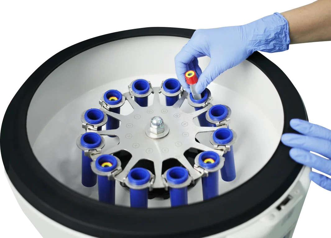
Nuclear Medicine
Nuclear Medicine is a branch of medical imaging, which uses a small amount of radioactive substance injected into the body, inhaled or swallowed, to diagnose and treat a variety of diseases including cancers, gastrointestinal, heart disease, endocrine and neurological disorder. It is often able to identify abnormalities very early in the progression of a disease, long before many medical problems are apparent with other diagnostic tests.
After diagnosis, and when treatment starts, these scans continue to play an important role in monitoring its effects and progress of treatment regime, to determine if modification of therapy is required.
Established in 2018, the Advance Nuclear Diagnostic and Therapeutic Centre @ PHKL is equipped with cutting-edge advanced technology in Nuclear Medicine (PET-CT & SPECT-CT) to deliver accurate structural and functional imaging of the body for diagnosis and management of cancer and other diseases. This allows for tailored individualised treatment plans for our patients, which translate to better prognosis and outcomes for our patients.
Biograph mCT S 64 PET-CT
READ MORE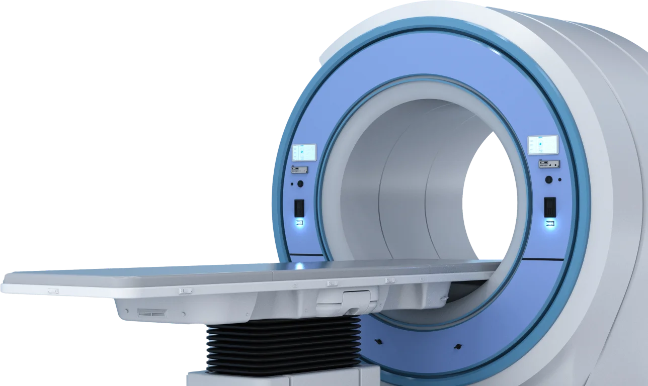
Symbia Intevo
T16 SPECT-CT
READ MORE


.webp?sfvrsn=c12d7068_11)

Siemens Biograph mCT S 64 PET-CT
The Siemens Biograph mCT S 64 is a whole-body PET/CT tomograph machine designed for the purposes of oncological, neurological and cardiac imaging and diagnosis. This machine has high spatial slice resolution and high sensitivity detector for faster acquisition. It is also capable of scanning the entire body in one scan with a long scan range of 203cm. With TrueV technology, it widens the axial field of view (FOV) and increases count rate performance. This enables scan time to be shortened for a faster imaging process.
The CT imaging capability of the Biograph mCT consists of a 64 slice CT. Thus, it offers full range spiral CT application. The machine also supports advanced RT simulation to further enhance accurate localisation of tumours. This advanced technology utilises fluorodeoxyglucose (F-18 FDG) to visualise the molecular function of active diseased cells, cancer in particular and its spread throughout the whole body. PET-CT is commonly used in cancer diagnosis as to its capability improving the cancer staging accuracy thus becoming the best modality in monitoring the progress of cancer treatment. The device is also proven to benefit patients diagnosed with Coronary Artery Diseases and with peripheral arteries including dementia especially Alzheimer Disease. The New 4-D PET-CT technology with respiratory gating allows an accurate assessment for treatment.
Besides the F-18 FDG, the machine also performs Ga-68 DOTATATE and PSMA PET-CT Scan – a radiopharmaceutical tracer used during PET Scans for prostate cancer and neuroendocrine tumour respectively. The Ga-68 DOTATE PET-CT full-body scan can capture neuroendocrine tumours (NETs) that overexpress somatostatin receptors and show where the tumours are located in the body. Ga-68 PSMA PET-CT full-body scan is a modern diagnostic imaging procedure and is becoming the gold standard in the diagnosis of prostate cancer. It is able to bind prostate tumour cells to be imaged using the PET technique. This technique also helps to monitor prostate cancer metastasis and recurrence even at low levels of PSA.

Siemens Symbia Intevo T16 SPECT-CT
The Siemens Symbia Intevo T16 SPECT/CT is a combination of state-of-the-art Single Photon Emission Computed Tomography (SPECT) and high quality 16 slices diagnostic Computed Tomography (CT). With xSPECT technology, true integration of SPECT and CT images are enabled. Together with a HD detector, it allows SPECT information to be registered into CT frame of reference resulting in higher SPECT images resolution and accurate, reproducible quantitative results. This machine can operate as SPECT-only, SPECT-CT or stand-alone CT diagnostic application.
The role of SPECT in showing biological functions through radio-tracer distribution in the body and when combined with CT modality showing precise location and anatomical structures making this advanced technology a superior diagnostic imaging modality than other conventional diagnostic imaging. The functional imaging produced by SPECT-CT includes whole body bone scintigraphy, thyroid scintigraphy, parathyroid scintigraphy, myocardial perfusion and heart function scintigraphy, gastrointestinal (GIT) bleed scintigraphy, renal scintigraphy, hepatobiliary scan, lung scintigraphy, brain scintigraphy, etc.
Why Choose Us?

450
Licensed
Beds

30+
Treatment
Modalities

26
Chemo
Bay

38
Medical
Specialities

200+
Specialist
Doctors

30,000+
Patient
Served
Patient Journey


Chemotherapy
Chemotherapy uses drugs to kill or slow the growth of cancer cells. It can be administered systemically (through the bloodstream) to target cancer cells throughout the body. Chemotherapy is used in various cancers, either as the primary treatment or in combination with other therapies, such as with surgery or radiotherapy.